Auditory Nerve Compound Action Potential During Intraoperative Auditory Nerve Monitoring
In cerebellopontine angle (CPA) surgeries, brainstem auditory evoked potentials (BAEP) have become a routine monitoring tool to assess the patient's auditory pathway and brainstem function. However, conventional BAEP monitoring requires a significant amount of signal averaging and has limited noise rejection capabilities. Today, we will explore an alternative monitoring technique - compound action potentials of the auditory nerve.At NCC, we are a leading provider of intraoperative neurophysiological monitoring solutions.
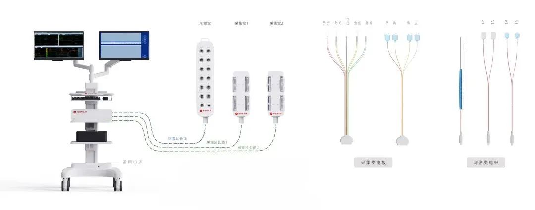
Compound action potentials of the auditory nerve, also known as "CAP potentials," directly record the compound action potentials of the eighth cranial nerve. The recording electrode, a cotton wick electrode, is placed on the exposed portion of the auditory nerve within the skull, while the reference electrode is positioned at the vertex (Cz). This method allows for the detection of functional activity in two different parts of the auditory nerve - near the brainstem and near the internal auditory canal. The recorded action potentials have high amplitudes and require fewer signal averages (around 10-15) to obtain a clear waveform.
Monitoring Parameters
Recording sites: A1/A2-Cz (brainstem auditory) Wick-Cz (NAP)
Analysis time: 15 ms
Sweep speed: 1.5 ms/div
Averaging: 1000 sweeps
Bandpass filters: 10-30 Hz (low-cut) to 2500-3000 Hz (high-cut)
In case of persistent electrical interference, the filter range can be adjusted to 100-200 Hz (low-cut) to 1000-1500 Hz (high-cut).
Stimulation parameters:
1. Stimulated side: 70 dB SPL, Contralateral side: Noise, 40 dB SPL
2. Stimulation rate: 11.1 Hz-15.9 Hz
3. Pure tone frequency: 1000-2000 kHz
Principles of Auditory Nerve CAP Recording
The principle behind recording auditory nerve compound action potentials using a wick electrode is straightforward. When sound stimuli enter the external auditory canal, pass through the tympanic membrane and middle ear, and reach the cochlea, the cochlea transforms the sound energy into neural action potentials. These action potentials then travel through the auditory nerve into the brainstem, bypassing the tumor. By placing the wick electrode near the brainstem side of the tumor, we can directly record the action potentials from the stimulated auditory nerve. Since the electrode is positioned close to the electrically active eighth cranial nerve, the response amplitude is typically higher than the normal BAEP wave I and can be obtained with fewer signal averages.
The main limitation of this method is that surgical manipulations, such as tumor exposure, traction, and resection, can cause the wick electrode to move away from the auditory nerve, resulting in a weakened or absent response signal. If the signal suddenly disappears, the surgeon should be notified to readjust the electrode position.
Recording Technique
The recording montage is similar to the standard BAEP recording setup, as auditory nerve CAP recording is performed after the tumor is exposed during surgery. First, BAEP is recorded using the conventional method. Once the tumor is exposed, the surgeon is asked to place the wick electrode between the tumor and the brainstem, where the auditory nerve enters the brainstem. To begin recording auditory nerve CAP, the ipsilateral earlobe needle electrode is replaced with the wick electrode, changing the montage from A1-Cz to wick electrode-Cz.
At NCC, we are a leading provider of intraoperative neurophysiological monitoring solutions. Our advanced equipment and dedicated support team ensure that surgeons have the tools they need to perform safe and effective procedures while minimizing the risk of neurological complications. If you're interested in learning more about our auditory nerve monitoring capabilities or any of our other products, please don't hesitate to contact us. Together, let's push the boundaries of what's possible in intraoperative neurophysiological monitoring.

 中文
中文 Arabic
 Spanish
 Hindi
Hindi French
 Indonesian
 Portuguese
 Persian
 Russian
 Korean
 German
 Vietnamese
 Turkish