中文
中文 Arabic
 Spanish
 Hindi
Hindi French
 Indonesian
 Portuguese
 Persian
 Russian
 Korean
 German
 Vietnamese
 Turkish
Clinical Solutions
Contact Us for Service & Support
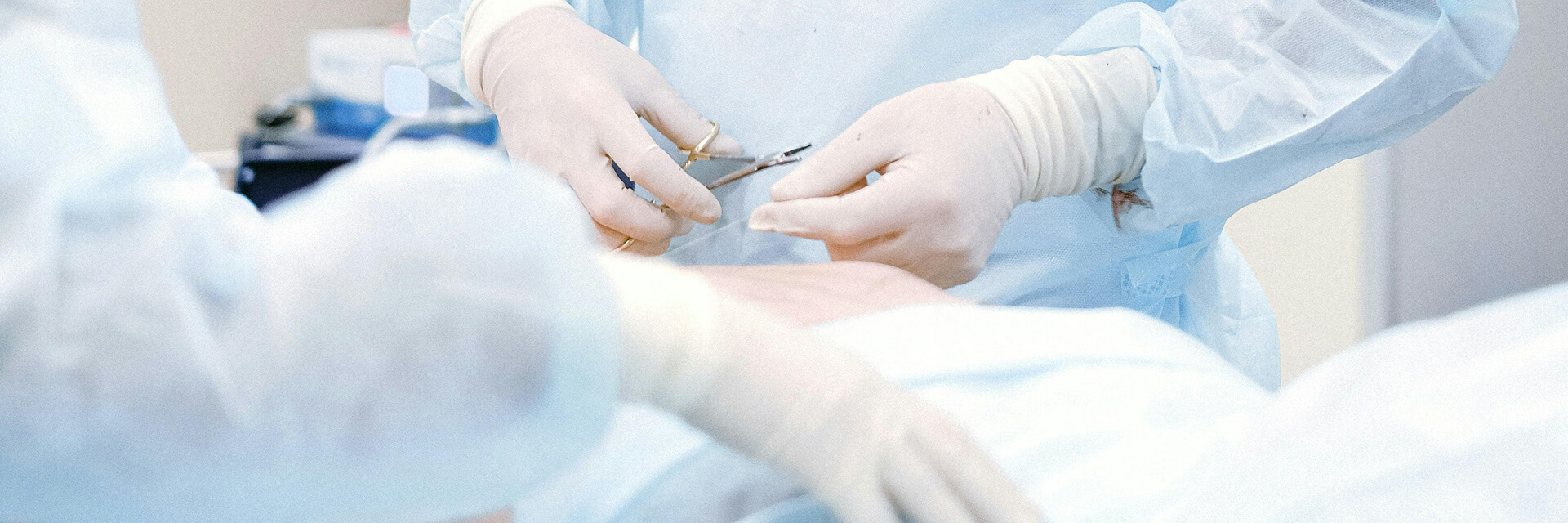
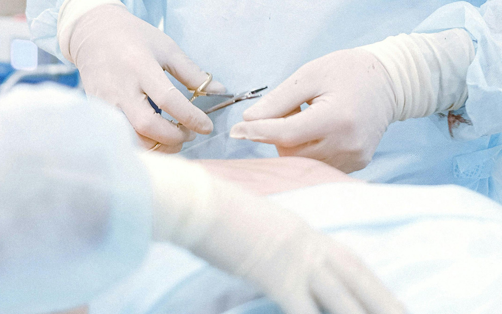
Orthopedic surgery(intraoperative neuromonitoring)
Cynapse IONM reduce risk in procedures including:
Cervical spine surgery
Thoracolumbar spine surgery
Coccyx surgery
Scoliosis correction surgery
Cervical spine surgery
Thoracolumbar spine surgery
Coccyx surgery
Scoliosis correction surgery
Key Features:
● User-friendly interface, easy to operate for neurophysiologist;
● Comprehensive monitoring of functional nerves at risk during surgery can be done simultaneously in various modes on a single screen, which can be switched freely;
● Modules can be monitored in various modes, edited and saved as templates for future use.
● Able to create and compare waveform before and after operations for better analysis.
● Real-time monitoring software displays real-time muscle relaxation, signal calibration, and interference adjustment during operations.
● The screen printing function allows for easy capture of monitoring waveform and automatic importation into reports or saving as pictures.
 390Electrical safety monitoring
 101Performance indicator testing
 4919Software testing and detection
 14Electromagnetic compatibility (EMC) testing
 6Biocompatibility testing
 8Simulated transportation test
Application
Orthopedic Intraoperative Neurophysiological Monitoring Solutions
Cervical Spine Surgery ▼
Somatosensory Evoked Potential (SSEP)
- Real-time monitoring of the ascending spinal pathways to protect the patient's sensory function.
- Stimulating Site: Median nerve or ulnar nerve (upper limbs), posterior tibial nerve (lower limbs).
- Recording Site: C3’-Fz, C4’-Fz (upper limbs), Cz-Fz (lower limbs).
Transcranial Electrical Motor Evoked Potential (TCe MEP)
- Real-time monitoring of the descending spinal pathways to assess and evaluate the patient's swallowing and motor function.
- Stimulating Site: C3, C4 or C1, C2 (precentral gyrus).
- Recording Site: Vocal cords (intubation electrodes), deltoid, abductor pollicis brevis, adductor hallucis, etc.
Free-run Electromyography (Free EMG)
- Observes real-time nerve traction responses.
Train-of-Four (TOF) Test
- Immediate monitoring of the patient's neuromuscular blockade status.
Thoracolumbar Surgery ▼
Somatosensory Evoked Potential (SSEP)
- Real-time monitoring of the ascending spinal pathways to protect the patient's sensory function.
- Stimulating Site: Posterior tibial nerve (lower limbs).
- Recording Site: Cz-Fz (lower limbs).
Transcranial Electrical Motor Evoked Potential (TCe MEP)
- Real-time monitoring of the descending spinal pathways to assess and evaluate the patient's motor function.
- Stimulating Site: C3, C4 or C1, C2 (precentral gyrus).
- Recording Site: Abductor pollicis brevis, adductor muscles, rectus femoris, tibialis anterior, abductor hallucis.
Free-run Electromyography (Free EMG)
- Observes real-time nerve traction responses.
Pedicle Screw Testing
- Automatically detects the accuracy of each screw placement.
Train-of-Four (TOF) Test
- Immediate monitoring of the patient's neuromuscular blockade status.
Scoliosis Orthopedics Surgery ▼
Somatosensory Evoked Potential (SSEP)
- Real-time monitoring of the ascending spinal pathways to protect the patient's sensory function.
- Stimulating Site: Median nerve or ulnar nerve (upper limbs), posterior tibial nerve (lower limbs).
- Recording Site: C3’-Fz, C4’-Fz (upper limbs), Cz-Fz (lower limbs).
Transcranial Electrical Motor Evoked Potential (TCe MEP)
- Real-time monitoring of the descending spinal pathways to assess and evaluate the patient's motor function.
- Stimulating Site: C3, C4 or C1, C2 (precentral gyrus).
- Recording Site: Abductor pollicis brevis, rectus abdominis, adductor muscles, rectus femoris, tibialis anterior, abductor hallucis, anal sphincter.
Free-run Electromyography (Free EMG)
- Observes real-time nerve traction responses.
Pedicle Screw Testing
- Automatically detects the accuracy of each screw placement.
Train-of-Four (TOF) Test
- Immediate monitoring of the patient's neuromuscular blockade status.
Sacral and Coccygeal Bone Surgery ▼
Somatosensory Evoked Potential (SSEP)
- Real-time monitoring of the ascending spinal pathways to protect the patient's sensory function.
- Stimulating Site: Posterior tibial nerve (lower limbs).
- Recording Site: Cz-Fz (lower limbs).
Transcranial Electrical Motor Evoked Potential (TCe MEP)
- Real-time monitoring of the descending spinal pathways to assess and evaluate the patient's motor function.
- Stimulating Site: C3, C4 or C1, C2 (precentral gyrus).
- Recording Site: Abductor pollicis brevis, adductor muscles, rectus femoris, tibialis anterior, abductor hallucis, anal sphincter.
Triggered Electromyography (Trigger EMG)
- Identifies and differentiates nerves from unknown tissues.
Free-run Electromyography (Free EMG)
- Observes real-time nerve traction responses.
Bulbocavernosus Reflex (BCR) Monitoring
- Immediate monitoring of the patient's sacral nerve reflex.
Train-of-Four (TOF) Test
- Immediate monitoring of the patient's neuromuscular blockade status.
Peripheral Nerve Surgery ▼
Nerve Conduction Velocity (NCV)
- Nerve conduction velocity is a diagnostic technique used to evaluate the conduction function of peripheral nerves, typically including motor nerve conduction velocity (MCV) and sensory nerve conduction velocity (SCV).
- Abnormalities in MCV and SCV are manifested as slowed conduction velocity and reduced amplitude. NCV measurement can be used to identify and differentiate peripheral nerves during surgery.
Somatosensory Evoked Potential (SSEP)
- Real-time monitoring of the ascending pathways to protect the patient's sensory function.
- Stimulating Site: Median nerve or ulnar nerve (upper limbs), posterior tibial nerve (lower limbs).
- Recording Site: C3’-Fz, C4’-Fz (upper limbs), Cz-Fz (lower limbs).
Transcranial Electrical Motor Evoked Potential (TCe MEP)
- Real-time monitoring of the descending pathways to assess and evaluate the patient's motor function.
- Stimulating Site: C3, C4 or C1, C2 (precentral gyrus).
- Recording Site: Corresponding muscles of the surgical limb.
FAQ
1. Intraoperative Neurostimulation Monitor: An Innovative Solution in the Field of Intraoperative Neuroelectrophysiological Monitoring ▼
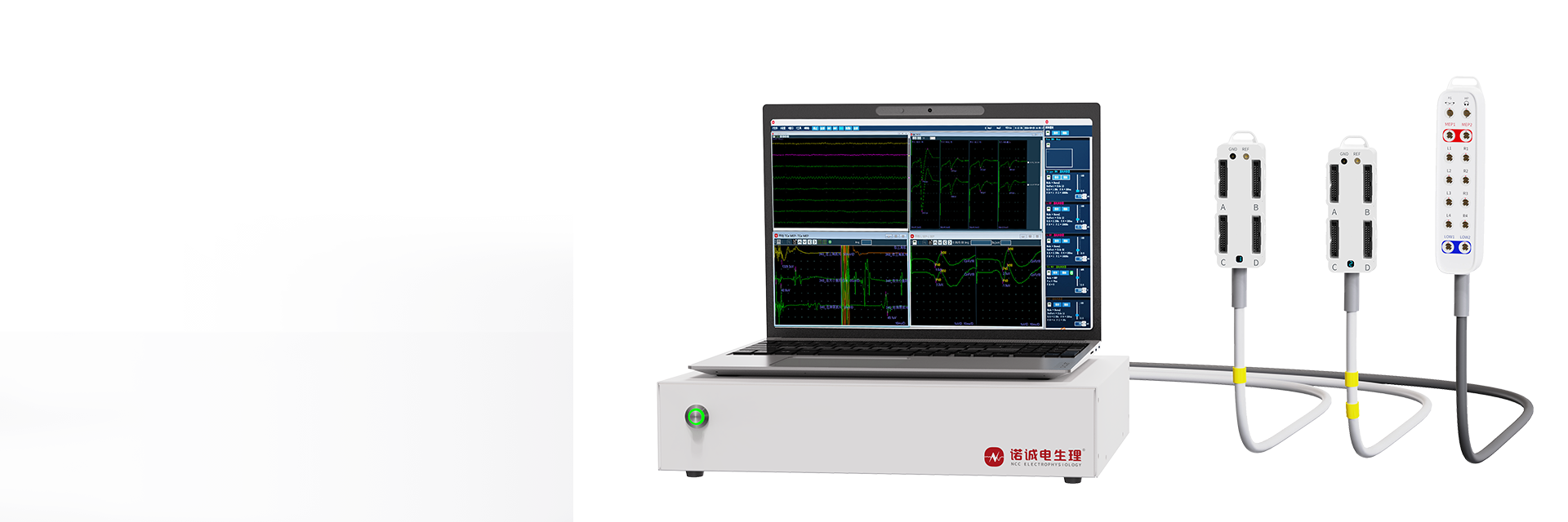
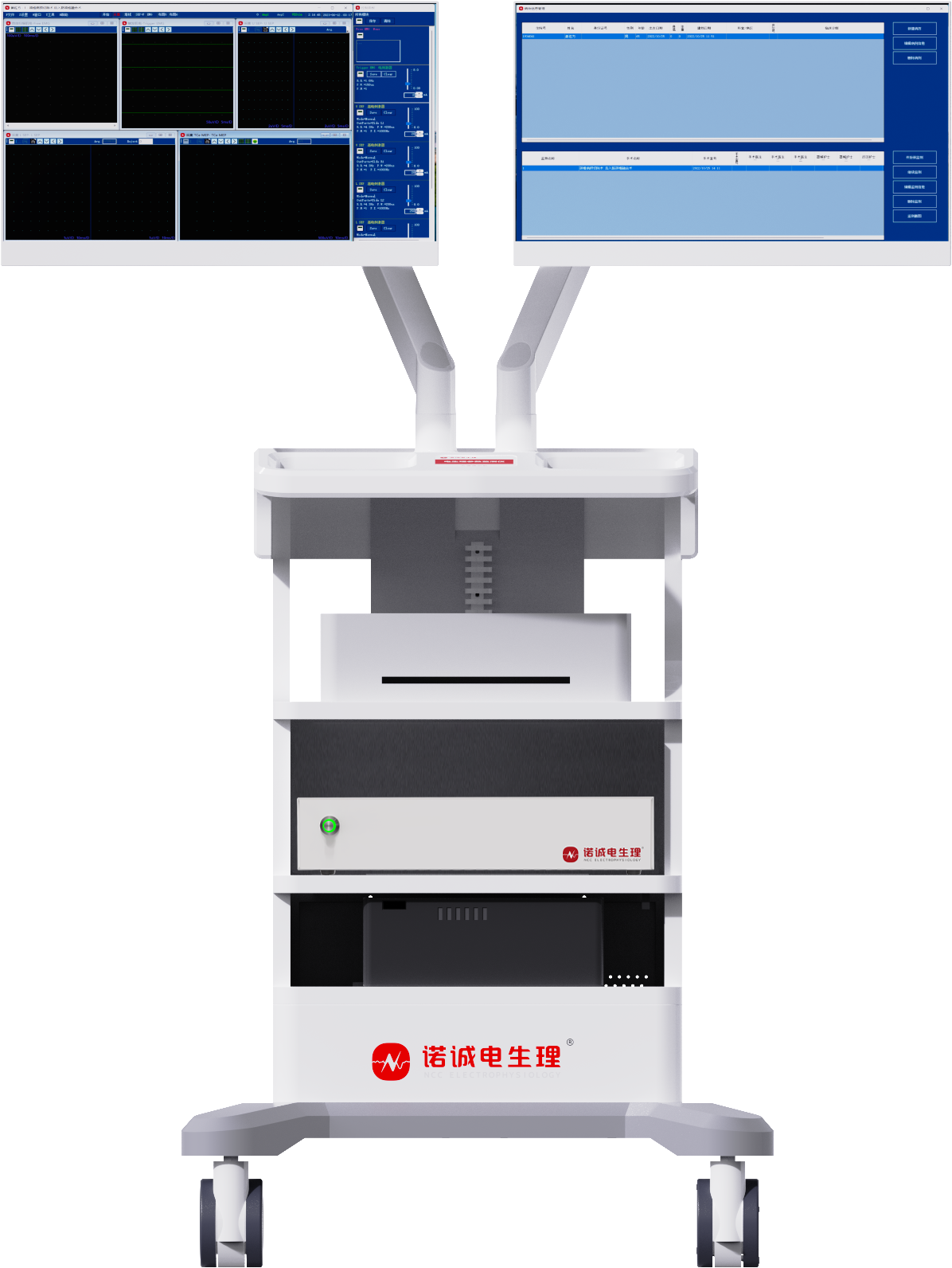
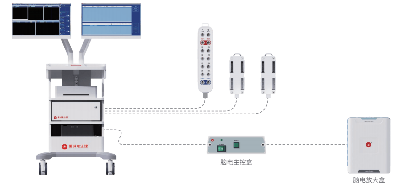
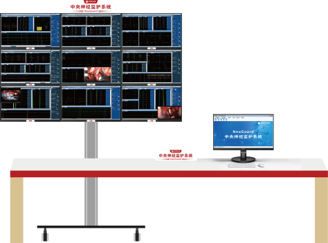
The intraoperative neurostimulation monitor developed by Nuocheng Electrophysiology is China's first intraoperative neuroelectrophysiological monitoring device applicable to complex surgical procedures and has obtained NMPA Class III medical device certification. The device provides monitoring of 32-channel IONM (Intraoperative NeuroMuscular Monitoring) + 256-channel EEG. It supports dual-screen adjustable display, with the main screen for real-time monitoring and the auxiliary screen synchronized with a microscope or video for convenient surgical operation.
The device has high-performance data acquisition capabilities, supports a portable design, and is equipped with all accessories for easy portability.
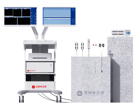
Figure: Intraoperative Neurostimulation Monitor (Dual-Screen)
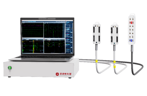
Figure: Intraoperative Neurostimulation Monitor (Portable)
2. Multi-module Monitoring ▼
Supports multi-module monitoring, including SEP (Somatosensory Evoked Potential), MEP (Motor Evoked Potential), EMG (Electromyography), BCR (Bulbocavernosus Reflex), Motor Cortex Localization, Language Cortex Localization, EEG (Electroencephalography), BAEP (Brainstem Auditory Evoked Potential), VEP (Visual Evoked Potential), Blink Reflex, D-Wave, Pedicle Screw Monitoring, and other 16 monitoring modules.

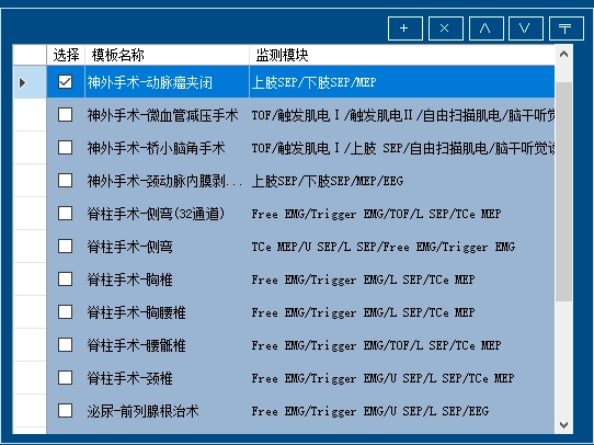
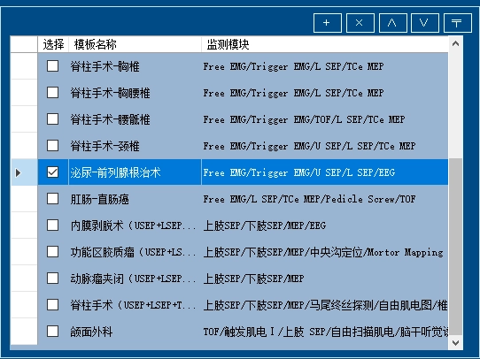
3. Function of personalized custom editing and saving of surgical procedures ▼
It contains more than 20 templates for various surgical procedures, which can be personalized and saved as surgical templates according to monitoring needs.
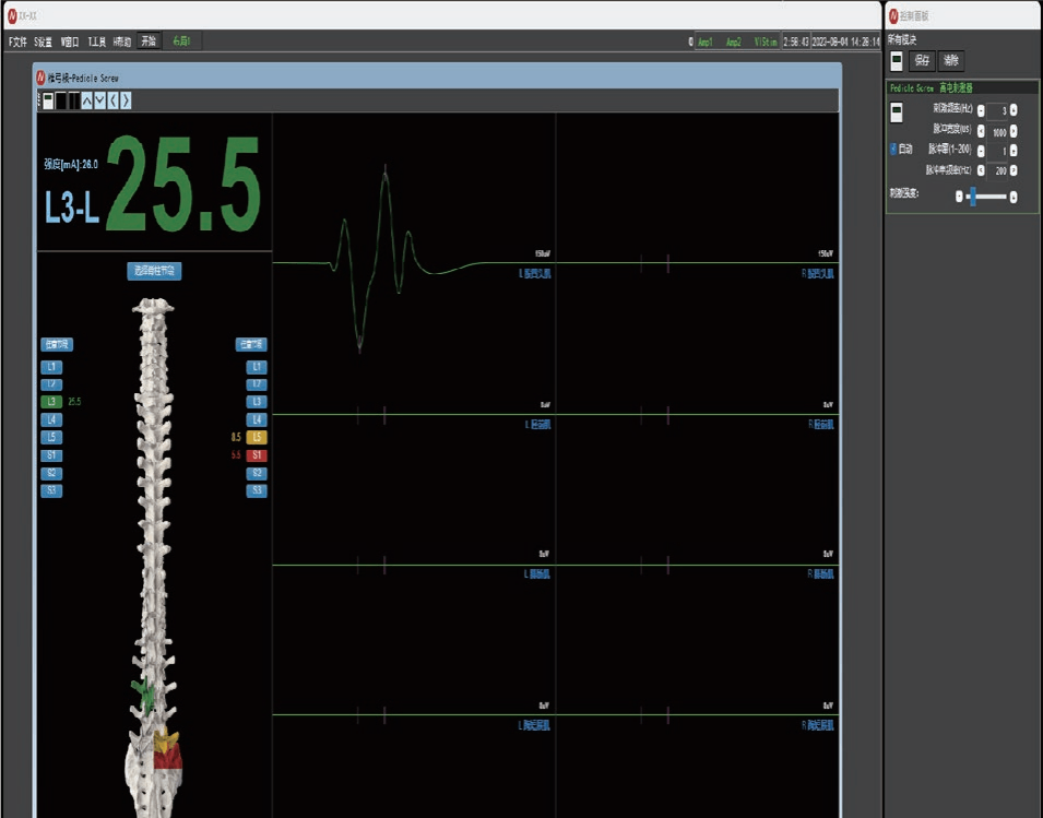
4. Spine Surgery - Automated Pedicle Screw Monitoring Module ▼
Automatically applies incremental pedicle probe currents. When the threshold is reached, the system stops intraoperatively without manual intervention, thereby monitoring whether the pedicle bone wall is breached and alerting the surgeon to avoid injuring adjacent nerve roots.
5. Neurosurgery - Functional Linkage between Intraoperative Neurophysiological Monitoring Technology (INOM) and 256-channel EEG Monitoring Technology ▼
• The high-throughput signals from the 256-channel EEG provide a data foundation for brain-computer interface (BCI) development, while INOM verifies the accuracy of neural signal decoding in real time. Together, they enable breakthrough experiments such as "thought dialogue," accelerating the clinical translation of "brain-controlled" intelligent devices and postoperative rehabilitation.
• By integrating technologies, the precision of neurosurgical procedures is enhanced. In the future, combined with artificial intelligence, deeper breakthroughs are expected in the fields of precise epilepsy treatment, brain function protection, and personalized medicine.
6. The Global First-In-Class Intraoperative Neurostimulation Monitoring Device - NeuGuard Central Neurological Monitoring System ▼
Cross-regional real-time collaboration hub, global technology resource sharing ecology
• to establish the world's first open neuroelectrophysiological collaboration platform to support expert consultation between multinational medical institutions (for example, Australian experts provide expert advice to Indonesian surgeons remotely)
• to achieve the efficient allocation of experienced electrophysiologists resources and break geographical barriers and to establish a 24-hour expert support network
• Provide standardized neuromonitoring support to medically underserved areas, such as hospitals in remote, underdeveloped regions or island-based medical facilities
• junior operators can get real-time expert guidance through the system, reducing the dependence on highly experienced technicians
• The NeuGuard Central Neurological Monitoring System breaks through the traditional "one-to-one" monitoring mode, enabling a single technician to simultaneously and precisely manage multiple surgeries
7. Innovative Breakthrough in Consumables - Rainbow Electrode ▼
• All-in-one design, easy operation
• The operation can be completed in one connection, greatly improving the efficiency.
• Guide the needle by color, intuitive and easy to understand, quick to use.
• Customized consumables combination, flexible adaptation
• Clinical-oriented consumable package design
• To meet the diversified needs of surgery, a special consumable package is provided for different surgical procedures.
• Strong anti-interference ability, accurate and reliable data
• Integrated consumables with excellent anti-interference performance, significantly improve the waveform fidelity.
• It provides more accurate and reliable monitoring data for surgery, and helps to ensure the safety and success of surgery.
NCC Rainbow electrodes-simplified operation, accurate monitoring, Safeguarding surgery!
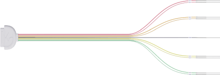
NCC Rainbow Electrodes
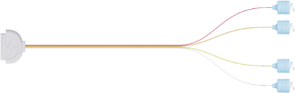
8. Innovative Breakthrough in Consumables - Radiolucent Electrode ▼
• Radiolucent Technology: Utilizes advanced carbon fiber material, which is invisible under DSA, reducing interference during surgery.
• Enhance Surgical Precision: Eliminate imaging interference, allowing surgeons to focus more on the surgical procedure and improving surgical success rates.
• Material Innovation: The application of carbon fiber material, which is lightweight and has high strength, provides stable support for surgery.
• Exceptional Performance: Stronger anti-interference capability and faster transmission rate ensure the stability and real-time performance of signals during surgery.